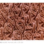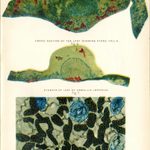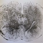
Lower Epidermis of Leaf Showing the Stomata and Chlorophyll Cells [Fig. 1]-
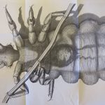
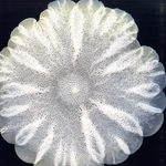
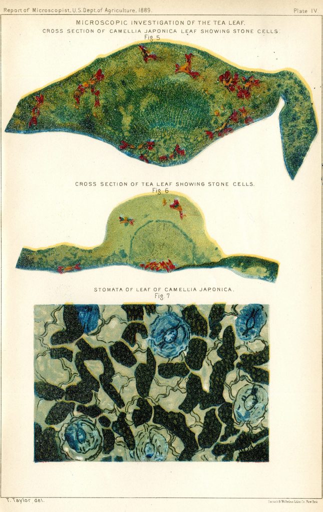
Microscopic Investigation of the Tea Leaf
Illustrated by T. Taylor
Printed by Sackett & Wilhelms Lith Co., New York
First Report of the Secretary of Agriculture- 1889
Washington: Government Printing Office, 1889
Gould Library Government Documents
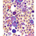
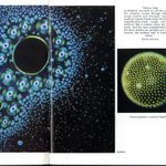
The books on display here, all from the collections of Gould Library, reveal the beauty and order of hidden worlds. In these volumes, microscopic images are deployed for a variety of reasons: Sometimes their purpose is purely practical (to certify the purity of a sample of tea, or to facilitate the identification of diseased blood). In other cases, as in Felice Frankel’s close-up view of Velcro, their job is as much to inspire curiosity and wonder as it is to illustrate a scientific phenomena.